Myocarditis Ecg / Pem Sim Case 1 Ecg Myocarditis Aliem / 23 ecg abnormalities, however, are widely variable, and there is not one specific abnormality that occurs with enough frequency to be a specific marker.
Myocarditis Ecg / Pem Sim Case 1 Ecg Myocarditis Aliem / 23 ecg abnormalities, however, are widely variable, and there is not one specific abnormality that occurs with enough frequency to be a specific marker.. Ecgs can help to identify myocarditis, although the findings are not specific. A diagnosis may be supported by an electrocardiogram (ecg), increased troponin, heart mri, and occasionally a heart biopsy. 23 ecg abnormalities, however, are widely variable, and there is not one specific abnormality that occurs with enough frequency to be a specific marker. An echocardiogram might detect enlargement of your heart, poor pumping function, valve problems, a clot within the heart or fluid around your heart. However, there are no data on their utility in ici myocarditis.
In the setting of normal ecg, troponin, and inflammatory markers, myocarditis or pericarditis are unlikely. Myocarditis is an inflammation of the heart muscle (myocardium). All our ecgs are free to reproduce for educational purposes, provided: Often associated with pericarditis, termed myopericarditis. The image is credited to litfl.com.
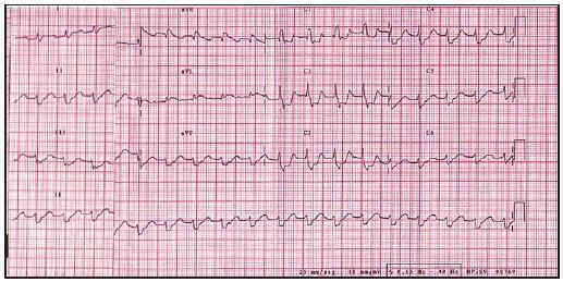
Giant Cell Myocarditis A Case Report Revista Espanola De Cardiologia from multimedia.elsevier.es Myocarditis has a number of etiologies. Bennett werner answered 44 years experience cardiology pretty much: Later on only t abnormalities, usually negative waves, are present and these occur in all. St elevation without reciprocal st depression was one of the conspicuous findings in the acute stage. 1 the etiology of myocarditis is heterogeneous but can be broadly categorized into infectious, toxic or autoimmune insults. All our ecgs are free to reproduce for educational purposes, provided: 23 ecg abnormalities, however, are widely variable, and there is not one specific abnormality that occurs with enough frequency to be a specific marker. There is no single diagnostic test in the ed that will confirm a diagnosis of myocarditis but several investigations will aid in making a clinical diagnosis.
The american heart association (aha) recommends further testing for patients having signs consistent with myocarditis with 1 or more cardiac imaging methods such as echocardiogram or cardiovascular magnetic resonance (cmr).10the echocardiogram usually is more readily deployed because it is portable.
The purpose of the present study was to clarify the characteristic findings of electrocardiogram (ecg) in 11 patients with acute myocarditis. Ecgs are virtually always abnormal in children with myocarditis, but a normal ecg does not rule out the possibility of the disease. The american heart association (aha) recommends further testing for patients having signs consistent with myocarditis with 1 or more cardiac imaging methods such as echocardiogram or cardiovascular magnetic resonance (cmr).10the echocardiogram usually is more readily deployed because it is portable. Myocarditis rests largely on the ecg findings and, of course, on the history. In mild cases, it may not show any deviation of waves. Myocarditis is most often due to a viral infection. Background myocarditis is a highly morbid complication of immune checkpoint inhibitor (ici) use that remains inadequately characterized. In the acute setting can cause arrhythmias, cardiac failure, cardiogenic shock and death. The image is credited to litfl.com. In the ed an ecg, chest radiograph (cxr), echocardiography and cardiac biomarkers should be performed at an early stage. 1 the etiology of myocarditis is heterogeneous but can be broadly categorized into infectious, toxic or autoimmune insults. If there is also pericarditis, there are subtle changes that can sometimes be seen.other tests can be done to diagnose myocarditis, including an echocardiogram and a cardiac mri. 23 ecg abnormalities, however, are widely variable, and there is not one specific abnormality that occurs with enough frequency to be a specific marker.
It was undetermined if these patients had myocarditis. Methods from an international registry, ecg parameters were. Background myocarditis is a highly morbid complication of immune checkpoint inhibitor (ici) use that remains inadequately characterized. Ecgs show only episodes of sinus tachy w/ normal resting rate. He was in nice, france, on holiday.
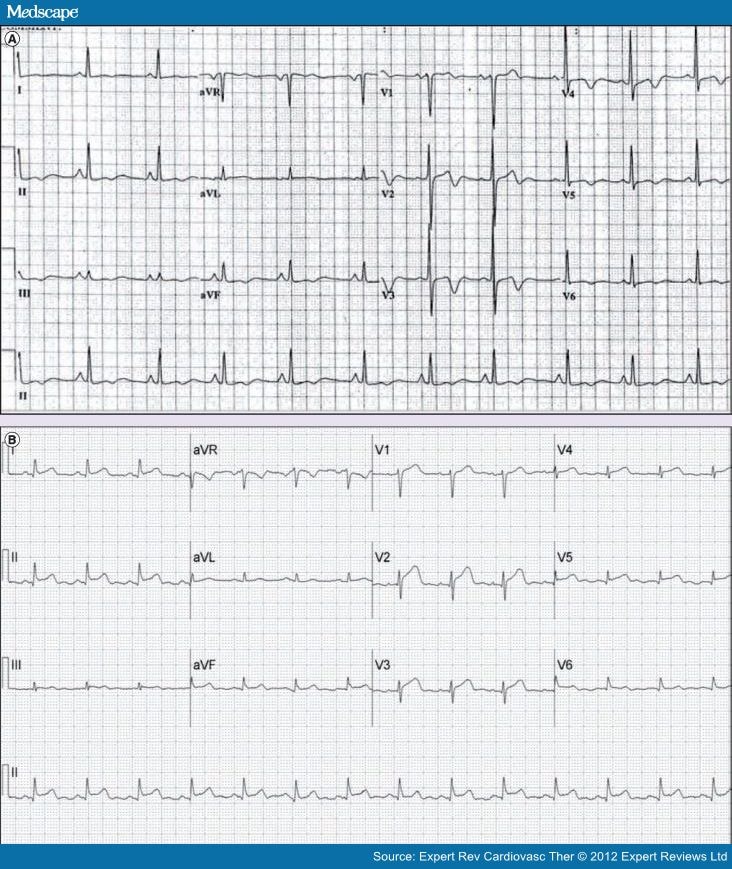
Endocarditis And Myocarditis from img.medscapestatic.com Background myocarditis is a highly morbid complication of immune checkpoint inhibitor (ici) use that remains inadequately characterized. Myocarditis is an inflammation of the heart muscle (myocardium). Ecgs show only episodes of sinus tachy w/ normal resting rate. An echocardiogram might detect enlargement of your heart, poor pumping function, valve problems, a clot within the heart or fluid around your heart. In the setting of normal ecg, troponin, and inflammatory markers, myocarditis or pericarditis are unlikely. Methods from an international registry, ecg parameters were. Inflammation from viral etiologies is thought to be caused both by direct cellular damage by the infectious agent and also from involvement by the host's immune system. Myocarditis can affect your heart muscle and your heart's electrical system, reducing your heart's ability to pump and causing rapid or abnormal heart rhythms (arrhythmias).
Viral myocarditis is the most common etiology in the developed world and the focus of this discussion.
He was in nice, france, on holiday. Nevertheless, ecg is widely used as an initial screening tool for myocarditis. If there is also pericarditis, there are subtle changes that can sometimes be seen.other tests can be done to diagnose myocarditis, including an echocardiogram and a cardiac mri. In myocarditis, ecg is an asset to find out abnormal heart rhythms. Myocarditis rests largely on the ecg findings and, of course, on the history. The purpose of the present study was to clarify the characteristic findings of electrocardiogram (ecg) in 11 patients with acute myocarditis. Myocarditis can be silent on an ekg. The qrs duration and the qtc interval are standardized electrocardiographic measures that are prolonged in other cardiac conditions; In the ed an ecg, chest radiograph (cxr), echocardiography and cardiac biomarkers should be performed at an early stage. Myocarditis is a disease marked by the inflammation of the heart muscle known as the myocardium — the muscular layer of the heart wall. It was undetermined if these patients had myocarditis. An echocardiogram might detect enlargement of your heart, poor pumping function, valve problems, a clot within the heart or fluid around your heart. The image is credited to litfl.com.
In the acute setting can cause arrhythmias, cardiac failure, cardiogenic shock and death. He was in nice, france, on holiday. Myocarditis is an inflammation of the heart muscle (myocardium). In such cases, it is advisable to avoid exercises and sports. In myocarditis, ecg is an asset to find out abnormal heart rhythms.
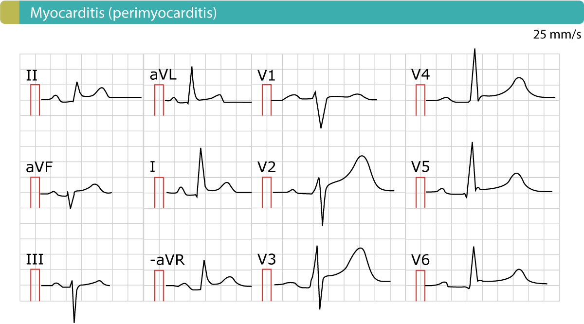
Dnbfrgbuxyglkm from ecgwaves.com Myocarditis can be silent on an ekg. In myocarditis, ecg is an asset to find out abnormal heart rhythms. This muscle is responsible for contracting and relaxing to. A diagnosis may be supported by an electrocardiogram (ecg), increased troponin, heart mri, and occasionally a heart biopsy. Myocardial inflammation in the absence of ischaemia. For suspected cases, consider consultation with cardiology for assistance with cardiac evaluation and management. Inflammation from viral etiologies is thought to be caused both by direct cellular damage by the infectious agent and also from involvement by the host's immune system. Methods from an international registry, ecg parameters were.
Ecgs can help to identify myocarditis, although the findings are not specific.
If there is also pericarditis, there are subtle changes that can sometimes be seen.other tests can be done to diagnose myocarditis, including an echocardiogram and a cardiac mri. Total qrs amplitudes at the acute stage were significant … 1 the etiology of myocarditis is heterogeneous but can be broadly categorized into infectious, toxic or autoimmune insults. Litfl ecg library is a free educational resource covering over 100 ecg topics relevant to emergency medicine and critical care. Myocarditis is an inflammation of the heart muscle (myocardium). Myocarditis can be silent on an ekg. Ecg changes in acute pericarditis, myocarditis, perimyocarditis the ecg is used to diagnose acute pericarditis. This muscle is responsible for contracting and relaxing to. Methods from an international registry, ecg parameters were. One must always rule out the most serious differential diagnosis, which is st elevation myocardial infarction (stem). Myocarditis has a number of etiologies. An echocardiogram might detect enlargement of your heart, poor pumping function, valve problems, a clot within the heart or fluid around your heart. Pericarditis produces st elevations at first, often in eleven of the twelve leads with st depression in the twelfth, avr.
Myocarditis is an inflammation of the heart muscle (myocardium) myocarditis. However, there are no data on their utility in ici myocarditis.


0 Komentar